
認知・情動脳科学専攻 Major of Cognitive and Emotional Neuroscience
Neurobiological study in psychiatric disorders
 神経精神医学 Neuropsychiatry
髙橋 努 Tsutomu Takahashi
- TEL : 076-434-7323
- URL : http://www.med.u-toyama.ac.jp/neuropsychiatry/index.html
- Keywords : Schizophrenia, Magnetic resonance imaging, Event-related potential, At-risk mental state, Bipolar disorder, Depressive disorder, Personality disorder.
研究の背景と目的 Background and Purpose of Study
統合失調症(schizophrenia)は思春期から青年期に好発する難治性精神疾患であり、その頻度はおよそ100人にひとりと決して稀ではない。遺伝的要因、脳内の神経伝達物質のアンバランス、環境要因などが統合失調症の発症に関連すると考えられるが、その神経生物学的基盤は不明な点が多い。磁気共鳴画像(magnetic resonance imaging, MRI)などを用いた脳画像研究により統合失調症における前頭-側頭-辺縁系領域を中心とした軽微かつ広範な灰白質体積減少などが報告されるが、これらの変化が生じるタイミング、疾患経過中の変化、疾患特異性についてはほとんどわかっていない。われわれのグループは、精神疾患の神経生物学的基盤の解明に向けて、統合失調症、精神病ハイリスク症例(at-risk mental state, ARMS)、その他の精神疾患 [双極性障害(bipolar disorder)、うつ病性障害(depressive disorder)、パーソナリティ障害(personality disorder)など] を対象にMRIや事象関連電位(event-related potential, ERP)など用いた生物学的研究を行っている。
Schizophrenia is a severe mental illness that generally appears in late adolescence or early adulthood with the prevalence of about 1% of adult population. Genetic and environmental factors as well as chemical imbalance in the brain may contribute to the development of schizophrenia, but its exact neurobiology remains largely unknown. Neuroimaging studies using magnetic resonance imaging have demonstrated subtle but widespread gray matter reduction predominantly in frontal and temporo-limbic brain regions in schizophrenia, but timing, course, and disease specificity of these morphologic changes have not been well documented. Our group has examined the characteristics of schizophrenia, at-risk mental state (ARMS), and other psychiatric disorders (e.g., bipolar disorder, depressive disorder, and personality disorder) using brain MRI, event-related potential (ERP), and other methodologies for a better understanding of the neurobiological basis of psychiatric disorders.
本研究の領域横断性
種々の精神疾患でみられる症状はヒトの認知、情動、行動およびそれらの異常に関するモデルとして有用であり、われわれの研究は基礎と臨床をつなぐトランスレーショナル研究や創薬研究において臨床サイドからのアプローチとして有用と考えられる。また精神疾患で認められる神経生物学的所見(脳形態変化など)は分子遺伝学的研究における中間表現型としての有用性が示されている。画像解析や神経生理学的研究においては、新規の工学技術や数学モデルを取り入れることで、さらに多くの臨床に有用な知見が得られることが期待できる。
研究内容
1)統合失調症早期の脳形態変化
統合失調症を対象としたMRI研究では、統合失調症患者には主に胎生期における神経発達障害を反映すると思われる粗大な脳形態の特徴(正中構造、脳溝脳回パターン、および大脳半球左右差の偏倚)(文献1)に加え、発症後に上側頭回などに進行性灰白質減少を認めた(文献2)。これらの進行性変化は臨床症状の重症度と相関し、また抗精神病治療により軽減されることが示唆された。これらの知見などから、統合失調症圏に推定される疾患経過中に生じる脳形態変化のモデルを提唱している(図1)。またコルチゾールを介したストレス反応の指標である下垂体体積は統合失調症で増大していることから、統合失調症発症におけるストレスの関与が示唆された(文献3)。
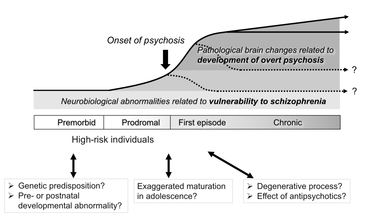
Figure 1. Hypothesized longitudinal brain morphologic changes in schizophrenia.
2)ハイリスク研究
閾値下の精神病様症状などから規定される精神病性障害の発症ハイリスク状態(at-risk mental state, ARMS)の症例では、1年以内に2-3割が統合失調症などの精神病性障害に移行すると報告される。われわれは、後に発症するARMS症例では発症に先立ち事象関連電位であるduration mismatch negativityの振幅減少がみられることを見出した(文献4)。またメルボルン大学との共同研究や国内多施設共同研究により、精神病性障害の発症前後において年間5%程度の比較的強い進行性萎縮が上側頭回にみられること(文献5, 図2)や後に発症するARMS症例では発症しない症例と比較して脳形態変化の程度が強いこと(文献6)などを明らかにした。
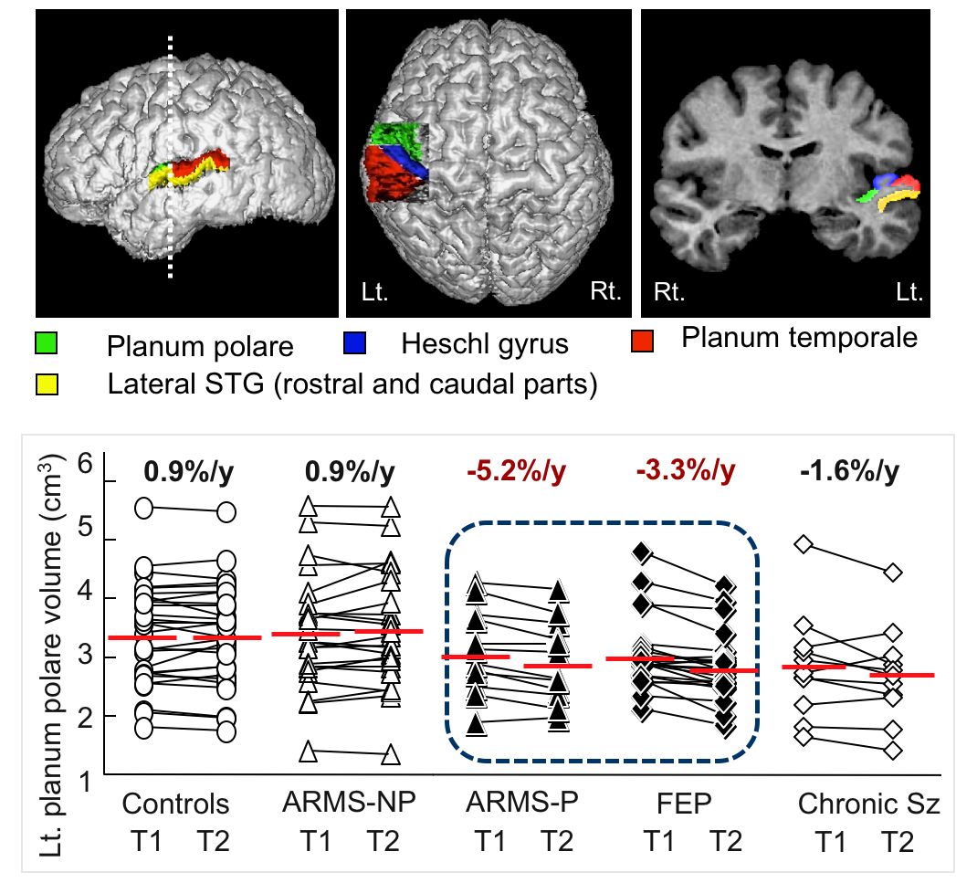
Figure 2. Longitudinal changes of the superior temporal gyrus in various stages of psychosis. FEP, first-episode psychosis; ARMS-NP, at-risk mental state without later psychosis onset; ARMS-P, at-risk mental state with later psychosis onset; Sz, schizophrenia; T1, time 1 (baseline scanning); T2, time 2 (follow-up scanning after approximately 2 years of baseline scanning).
3)臨床応用の可能性と課題
MRI画像を用いた判別分析により健常群と統合失調症群は8割程度の精度で判別可能であり(文献7)、生物学的指標による統合失調症の補助診断が期待される。統合失調症と双極性障害の脳形態を比較すると、各臨床病期における脳形態の特徴は異なっていたが、うつ病性障害やパーソナリティ障害では統合失調症と一部類似した脳形態変化を認めた。臨床応用に向けて、現在さらなる疾患特異性の検討および他の生物学的指標(ERP、嗅覚機能)のデータ蓄積を行っている。
参考文献
- Takahashi T, Takayanagi Y, Nishikawa Y, et al. Brain neurodevelopmental markers related to the deficit subtype of schizophrenia. Psychiatry Res Neuroimaging, 266: 10-18, 2017.
- Takahashi T, Suzuki M, Zhou SY, et al. A follow-up MRI study of the superior temporal subregions in schizotypal disorder and first-episode schizophrenia. Schizophr Res 119(1-3): 65-74, 2010.
- Takahashi T, Suzuki M, Velakoulis D, et al. Increased pituitary volume in schizophrenia spectrum disorders. Schizophr Res 108(1-3): 113-120, 2009.
- Higuchi Y, Sumiyoshi T, Seo T, et al. Mismatch negativity and cognitive performance for the prediction of psychosis in subjects with at-risk mental state. PLoS One 8(1): e54080, 2013.
- Takahashi T, Wood SJ, Yung AR, et al. Progressive gray matter reduction of the superior temporal gyrus during transition to psychosis. Arch Gen Psychiatry 66(4): 366-376, 2009.
- Sasabayashi D, Takayanagi Y, Takahashi T, et al. Increased occipital gyrification and development of psychosis in individuals with an at-risk mental state: a multicenter study. Biol Psychiatry, in press.
- Takayanagi Y, Takahashi T, Orikabe L, et al. Classification of first-episode schizophrenia patients and healthy subjects by automated MRI measures of regional brain volume and cortical thickness. PLoS One 6(6): e21047, 2011.